Close
The Vision Care Group of Companies has built a reputation as one of the best eye care solution providers in Sri Lanka by using an innovative approach and state-of-the-art equipment to ensure that customers receive world-class eye care at all branches. During our nearly 30 years of operations, we have successfully become the first company in Sri Lanka to bring the local eye care industry on par with global standards. We have also pioneered the introduction of the best range of eyewear brands recognized globally to the local market. Our high quality, high precision contact lenses, optical and ophthalmic products are available at all Vision Care outlets.
I am an IT Lead by profession. It’s a highly stressful job where I have to meet many top clients and impress them in order to win their trust, close sales and meet my targets. So, looking smart and dressing well is very important in my line of work. I needed a pair of top of the range branded spectacles and visited Vision Care. I was very impressed with their collection of luxury frames. The team was very helpful and gave me several options. I was able to get my specs pair in only a few days after ordering. Looking forward to visiting them again next year for my next pair!
Being a university student my budget is limited so I was worried about finding a good pair of specs. A friend told me to visit Vision Care because they have specs for all types of budgets. He was right. When I dropped in I saw a lot of options for my budget range. I was able to test my eyes then and there and also order a pair for a reasonable price. I am very happy with their staff and service. I will definitely return to Vision Care for my future specs needs.
As an entrepreneur working from my home office I am always running around meeting clients and also trying to get so many things done. I don’t have time to be visiting any showroom so I heard about the Vision Care Online Store and logged on to their site. The site was very nicely done and easy to use. I went through their large collection of sunglasses and found the perfect one, ordered, and paid online. The sunglasses came to my home in a few days and they are perfect. I am so happy at how easy the whole process was. I must say ordering online from Vision Care is the best option for busy people like me.
As a mother, buying anything for my daughter can be challenging as she is fussy sometimes. So, when the time had come to buy her a pair of spectacles, she insisted she wanted contact lenses and so I was expecting a tough time. We walked into our nearest Vision Care outlet. From the beginning, I was pleasantly surprised to see how they spoke with my daughter. They were very professional and courteous at all times. I am very thankful to Vision Care and its team for making this whole experience a wonderful one.
My father is very old and feeble and his eyesight is getting weaker. He has various health issues and is on different medications. I took him to Vision Care to buy his new pair of reading glasses as he couldn’t read properly with his old pair. He was his usual grumpy self but I was very happy to see that the staff there was very kind and patient with him. They answered all his questions and slowly took him through the entire process without agitating him. By the end of the visit, he was actually in a better mood and I was happy too. The glasses fitted him very well when we visited them a few days later.
Vision Care uses an innovative approach coupled with state-of-the-art equipment handled by our highly-qualified and well-experienced professionals to deliver a wide array of high quality services to all our valued customers across the island.


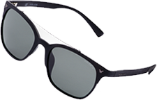
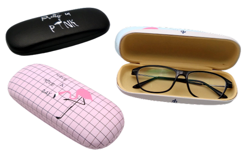
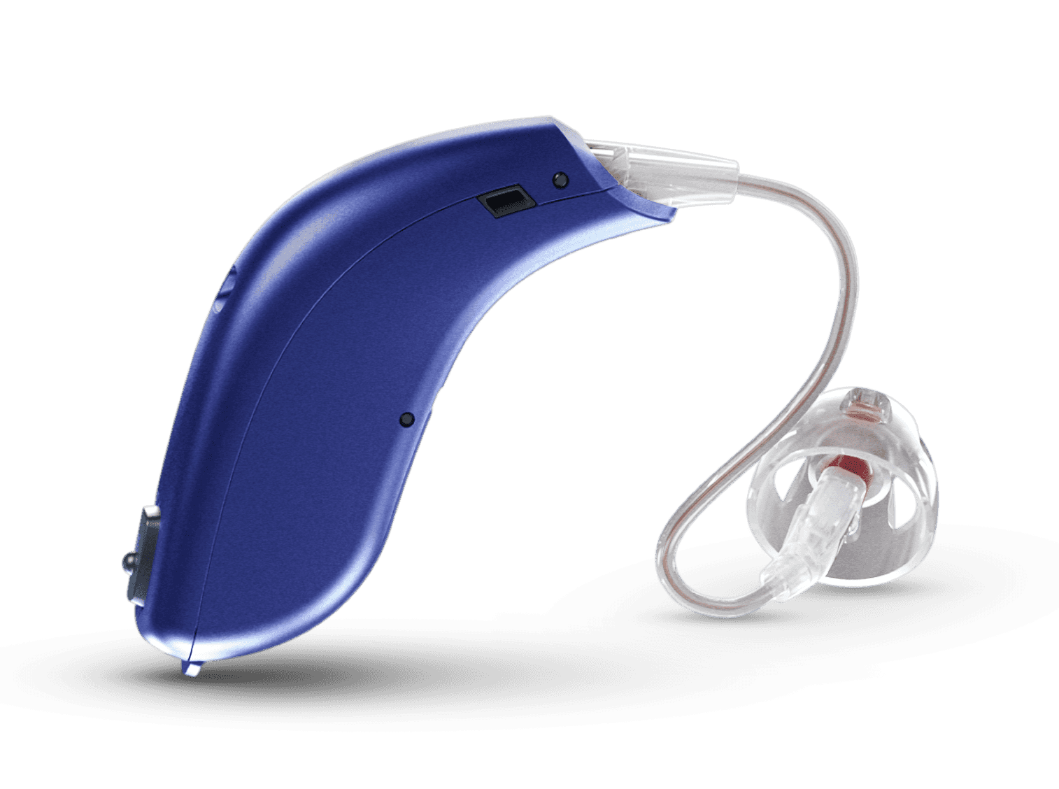
Find your perfect fit from the widest range of the world’s best eye care and hearing care brands available in Sri Lanka.
Take advantage of the latest credit and debit card discounts and offers from our banking partners
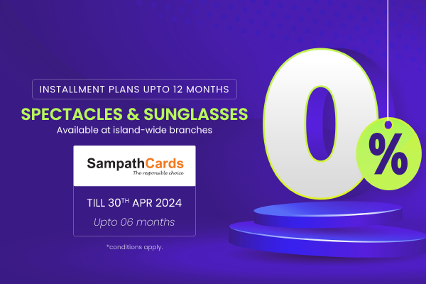
Experience the ease of Buy Now, Pay Later at Vision Care!
Purchase spectacles & sunglasses and enjoy installment options for up to 6 months with 0% interest when you use your Sampath Bank Credit Card.
Offer valid until April 30th at all Vision Care branches island-wide.
Terms and conditions apply.
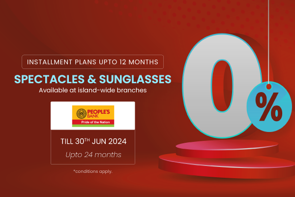
Shop at Vision Care with your People’s Bank Credit Card and enjoy 0% interest installments for 24 months on spectacles and sunglasses. Offer ends June 24.
T&C apply.

Vision Care, Sri Lanka’s largest vision, hearing and eyewear solutions provider, marked World Optometry Week with the launch of “Vision Care Primary Eye Screening Program,” as well as the introduction of the ‘Vision Care Eye Screening and Educational Toolkit’ This nationwide initiative aims to promote eye health awareness among school-aged children as well as private […]
Read MoreVision Care, Sri Lanka’s largest vision and eye wear solutions provider, celebrates International Women’s Day by dedicating a day to recognize the achievements and contributions of their female employees across all spectrums. Under the theme “Inspire Inclusion,” Vision Care reaffirms its commitment to fostering an environment where women from diverse backgrounds thrive and excel, empowering […]
Read MoreVision Care, Sri Lanka’s largest vision, hearing and eyewear solutions provider, extended its 10-year partnership with the Colombo Fashion Week (CFW) to its 11th year by once again collaborating as the “Official Eye and Hearing Care Partner” of CFW Summer 24 edition held recently. In a new initiative, Vision Care leveraged this platform for promoting […]
Read MoreStep into our newly established Vision Care Matara Branch! We’re thrilled to welcome you to our modern space at No. 161, Anagarika Dharmapala Mawatha, Matara. Our branch is thoughtfully designed for your comfort, featuring contemporary interiors and ample parking. Discover a wide array of high-quality optical and hearing services and products provided by our expert […]
Read MoreWelcome to our new Vision Care Ja-Ela Kapuwatte Branch! We are delighted to extend an invitation to our state-of-the-art facility, designed for your comfort and equipped with the latest in optical and hearing services and products. Conveniently located at 555C, Colombo Road, Kapuwatte, Ja-Ela, our modern branch boasts ample parking and a contemporary ambiance with […]
Read MoreStep into a world of elegance and innovation at the Vision Care Corporate Night held at our visionary 505 Branch on January 19, 2024. An enchanting evening where style met sophistication, as industry leaders and eyewear enthusiasts gathered to celebrate a fusion of cutting-edge eye care and timeless elegance. The ambiance sparkled with the glow […]
Read MoreVision Care celebrates a decade of glamour at Colombo Fashion Week as the “Official Eyewear Partner” 5 th December 2023, Colombo: In a dazzling display of fashion and vision, Vision Care, Sri Lanka’s largest vision and eyewear solutions provider, celebrated a decade of partnership as the "Official Eyewear Partner at Colombo Fashion Week’s Luxury Resortwear […]
Read MoreVision Care, Sri Lanka’s largest vision and eyewear solutions provider, successfully concluded the “Paata Siththam” 2023 Art Competition—an island-wide event crafted around the theme “Tomorrow’s World Through My Eyes.” This competition aimed to cultivate the creative talents of young artists in Sri Lanka, and the celebration of artistic expression took place in conjunction with World […]
Read More🖌️ලෝක ළමා දිනයට සමඟාමීව විෂන් කෙයා ආයතනය විසින් සංවිධානය කරනු ලබන දීප ව්යාප්ත චිත්ර තරඟාවලිය 2023 🎨📢 තරඟයේ තේමාව : ‘මගේ දෑසින් හෙට ලොව’ ඉදිරිපත් විය හැකි තරඟ අංශ : 👨🎨මූලික ප්රාථමික – 1,2,3 ශ්රේණි | මූලික ද්විතීක – 4,5,6 ශ්රේණි | කණිෂ්ඨ – 7,8,9 ශ්රේණි | ජේෂ්ඨ – 10, 11 ශ්රේණි | ජේෂ්ඨ ද්විතීක -12, 13 ශ්රේණි නිර්මාණ […]
Read MoreIt gives us great pleasure to announce that Vision Care, Sri Lanka’s largest vision and eye wear solutions provider, has expanded its islandwide network with the opening of its latest branch at Asiri Surgical Hospital, Colombo 05. Asiri Surgical Hospital is renowned for its specialized surgical services and advanced medical treatments, making it a fitting location […]
Read MoreVision Care, a leading eye care service provider, has expanded its reach with the opening of a new branch at Negombo Nawaloka Medicare premises on March 28th. The new branch will provide comprehensive eye care services to the local community, including eye exams, eyewear, and contact lenses. The opening of the new branch is part […]
Read MoreThe conveniently-located new Gampaha branch offers ample parking and a modern look and feel with spacious interiors and a wide range of products and services that includes spectacles, sunglasses, contact lenses and hearing aids. It is outfitted with state-of-the-art eye care and hearing care equipment that are expertly-handled by Vision Care’s well-trained, highly-experienced professionals. Customers […]
Read MoreWe’re excited to announce the opening of our newest branch at Wanduragala on February 16th, 2023. The Vision Care Wanduragala branch is a centrally-located, modern building with ample parking and spacious interiors. It provides a large collection of eye care products and services including spectacles, sunglasses, and contact lenses. Customers can experience the latest eye […]
Read MoreVision Care, Sri Lanka’s largest vision and eye wear solutions provider, has extended its Kurunegala branch by adding a state-of-the-art Eye and ENT Diagnostic Centre to provide customers with a wider selection of eye and hearing care solutions all under one roof. The new wing of the branch was officially declared open by Consultant Eye […]
Read MoreModel and lifestyle blogger Bishari needs sunglasses to protect her eyes when enjoying the great outdoors. Watch her as she visits the Racecourse Mall to take a look at what Vision Care, Sri Lanka’s largest vision and eye wear solutions provider, has to offer her. Vision Care brings you premium, luxurious international brands of eyewear […]
Read More𝑬𝒚𝒆𝒘𝒆𝒂𝒓 𝒇𝒐𝒓 𝒕𝒉𝒆 𝑺𝒆𝒂𝒔𝒐𝒏 𝒘𝒊𝒕𝒉 𝑽𝒊𝒔𝒊𝒐𝒏 𝑪𝒂𝒓𝒆 You can explore and experience the latest in the world of luxury eyewear at this branch from 10am to 10pm. For further details, call on 077 295 8487 or find us on maps: https://bit.ly/VCOneGalleFace Vision Care is Sri Lanka’s largest vision and eye wear solutions provider that brings […]
Read MoreDilmy is a university student studying hard for her upcoming exams. But her studies are disrupted as she finds it difficult to read her notes clearly without straining her eyes. She decides to stop studying and visit the branch of Vision Care, Sri Lanka’s largest vision and eye wear solutions provider, at the Colombo City […]
Read More𝐆𝐞𝐭 𝟏𝟏% 𝐨𝐟𝐟 𝐨𝐧 𝐒𝐩𝐞𝐜𝐭𝐚𝐜𝐥𝐞𝐬 𝐟𝐫𝐨𝐦 𝟏𝟓𝐭𝐡 𝐭𝐨 𝟑𝟏𝐬𝐭 𝐉𝐚𝐧𝐮𝐚𝐫𝐲 at our outlets in 𝗝𝗮𝗳𝗳𝗻𝗮, 𝗩𝗮𝘃𝘂𝗻𝗶𝘆𝗮, 𝗕𝗮𝘁𝘁𝗶𝗰𝗮𝗹𝗼𝗮, 𝗞𝗶𝗹𝗶𝗻𝗼𝗰𝗵𝗰𝗵𝗶, 𝗠𝗮𝗻𝗻𝗮𝗿, 𝗧𝗿𝗶𝗻𝗰𝗼𝗺𝗮𝗹𝗲𝗲 & 𝗪𝗲𝗹𝗹𝗮𝘄𝗮𝘁𝘁𝗲. Don’t miss out on this limited time offer, Visit us to Upgrade your look. #ThaiPongal #EyeWearOffer #Spectacles #Jaffna #Vavuniya #Batticaloa #Kilinochchi #Mannar #Trincomalee #Wellawatte #Sale #visionupgrade
Read MoreWhether you’re hitting the beach or running errands, these shades will keep you looking chic and on-trend. Vision Care offers a wide range of premium international brands of eyewear from across the globe. Friendly, experienced & qualified staff operate state-of-the-art equipment to provide you the best solutions for any eyewear requirement you may have. We […]
Read MoreVision Care offers a wide range of premium international brands of eyewear from across the globe. Friendly, experienced & qualified staff operate state-of-the-art equipment to provide you the best solutions for any eyewear requirement you may have. We have the ability to perform in-house eye examinations and provide the necessary testing for eyewear selection and […]
Read MoreVision Care Jaffna Branch incorporate with Veerakesari is delighted to inform you that the prize giving ceremony of Jaffna Art Competition held in commemoration of the World Children’s Day 2022 has been held on 15th November at Ilankaiventhan Kalai Kallury, Jaffna with great pomp and enthusiasm. 10 Winners have announced, over 500 participants in three […]
Read MoreThis World Sight Day, Let’s Raise Awareness For Better Eye Care For Everyone, Everywhere. Love Your Eyes – Protect & Make Your Eyes The Priority. Visit Our Islandwide Outlets, To Get Free Eye Checkups And Put Your Eye Health The Priority.
Read Moreවිෂන් කෙයා සහ නෙත් FM සංවිධානය කල “විශ්මලන්තය” ලෝක ළමා දින සැමරුම, කළුතර මහා විද්යාලයේදී සාර්ථකව පැවැත්විය. එහිදී කුඩා ළමුන් වෙනුවෙන් විශේෂ තරඟ රැසක් පැවැත්වූ අතර, ජයග්රහණය කළ කුඩා ළමුන් වෙනුවෙන් විෂන් කෙයා වෙතින් වටිනා ත්යාග ලබා දීමද සිදු විය.
Read MoreMovie Night 2022 Highlight Moments. Vision Care Movie Night 2022 took place last 30th September, which was organized by Vision Care Premium Outlet located at No 05 Ground Floor, Colombo City Centre. Here’s how it went!
Read MoreThe Free Health Camp organized by Badulla Vision Care Branch was held on September 25th at the branch premises. The camp was open to all who needed medical treatment for their health. Professional and qualified optometrists at Vision Care performed the eye examination and provided necessary guidance to the patients. Doctors checked blood pressure, blood […]
Read Moreවිෂන් කෙයා සහ නෙත් FM එක්ව සංවිධානය කරන 5 ශ්රේණිය ශිෂ්යත්ව සම්මන්ත්රණය සැප්තැම්බර් 18 වනදා, හෝමාගම මධ්ය මහා විද්යාලයේදී සාර්ථකව අවසන් විය. එහිදී පැමිණ සිටි දෙමාපියන් වෙනුවෙන් , දරුවන්ගේ අක්ෂි සෞඛ්ය සහ ආරක්ෂාව පිලිබඳ දැනුවත් කිරීමේ විශේෂ දේශනයක්ද පවත්වන්නට විෂන් කෙයා අපි කටයුතු කළා. එහිදී ශිෂ්යත්ව දරුවන් සහ දෙමාපියන් වෙනුවෙන් ත්යාග බෙදා දීම ද සිදු […]
Read Moreඔබ ලැබූ ජීවයට කළගුණ දැක්වීම ගුණය අපේ මුතුන් මිත්තන්ගෙන් අපට ලැබුණු අපුරු දායාදයක්. ලැබුවා වූ සිංහල හා දමිළ අලුත් අවුරුද්දේ සොබාදහමට පළමුව කළගුණ සැලකීම අමතක නොකළ විෂන් කෙයා ආයතනය, අපගේ පාරිභෝගිකයන් අතරෙන් පැළ බෙදාදීමේ වැඩසටහනක් සිංහල හා දමිළ අලුත් අවුරුද්දට සමගාමීව සිදු කරන ලදී. සුභ මොහොතට ඔබේ නිවසේ පැළයක් සිටුවා, මිහිකත සෙනෙහසින් සුරකින්න විෂන් කෙයා […]
Read MoreVision Care, Sri Lanka’s largest eye care solutions provider, has expanded its island wide network with the opening of its latest branch located at Ceymed Healthcare & Hospital Services, S.De.S Jayasinghe Mawatha, Kohuwala, Nugegoda. Ceymed Hospital is one of the leading hospitals in the Nugegoda region offering a wide range of healthcare services including eye […]
Read MoreVision Care celebrated International Women’s Day by dedicating a day to recognise, reward and educate their ‘visionary women’. To mark the occasion, Vision Care organised a training session, at the Vision Care Academy, where employees from its wide branch network were allowed to log in remotely. The training included an inspiring speech by Sri Lanka’s […]
Read MoreCelebrating 30 Years of Vision Care History and Staff Convention Ceremony – 2021 was held on 06th of February at Shangri-La Hotels with the participation of Vision Care all staff members. View Photos
Read MoreVision Care, Sri Lanka’s largest eye care solutions provider, declared open its newly-relocated Vavuniya branch recently. The new branch, conveniently-located at No. 52, Jaffna Road, Vavuniya, offers customers better park facilities, a modern look and feel, more interior space, and a wider range of products and services. As Sri Lanka’s No.1 eye care solutions provider, […]
Read MoreVision Care, Sri Lanka’s largest eye care solutions provider, unveiled its newly-revamped, more spacious Wellawatte outlet at its new location at No. 30, Galle Road, Colombo 06. The official opening ceremony was a low-key event held in accordance to the health guidelines given by the authorities. The relocation and reopening of the Wellawatte outlet is […]
Read MoreVision Care Academy, the educational institute of Sri Lanka’s largest eye care solutions provider Vision Care, celebrated its Optometry Day and Annual Scientific Session on 13th June with an event that was successfully carried out on a virtual platform in keeping with the health guidelines given out by the authorities. Held under the theme “Vision […]
Read MoreVision Care, Sri Lanka’s largest eye care solutions provider, has continued its commitment to boost sports in Sri Lanka by supporting the 105th Colombo Championship organised by the Sri Lanka Tennis Association (SLTA). The highly-anticipated tournament was held from 5th to 21st February 2021 with the participation of Sri Lanka’s finest tennis players across multiple […]
Read MoreVision Care, Sri Lanka’s largest eye care service provider, is marking World Sight Day 2020 under the global theme “Hope in Sight” by highlighting the importance of regular eye testing irrespective of a person’s age. Globally, as many as 1 billion people live with a preventable visual impairment or one that has yet to be […]
Read MoreVision Care, Sri Lanka’s largest eye care provider, has partnered with Colombo Fashion Week (CFW) once again as the “Fashionable Eyewear Partner” for the 2020 edition of the prestigious fashion event that was held across 3 days at the Hilton Colombo and Shangri-La Hotel. Vision Care collaborated with several designers including Emerging Designers Ayesh Wickramarathne, […]
Read Moreදිවයින පුරා සිටින අපගේ සියලුම ගනුදෙනුකරුවන්ට සුරක්ෂිත සේවාවක් සැපයීම සඳහා විෂන් කෙයා අපි උපරිම සෞඛ්යාරක්ෂිත පියවරයන් සමඟ දැන් සුදානම් වී සිටිමු. මේ පිළිබද ව සවිස්තරාත්මක රූපවාහිණී සාකච්ඡාමය වැඩසටහන.
Read MoreVision Care, Sri Lanka’s largest eye care provider, has opened all branches in the country and is ready to welcome customers while maintaining the highest standards in safety and hygiene in order to combat the spread of the COVID-19 disease. The company has introduced a number of safety measures to ensure the safety and bring […]
Read MoreVision Care, Sri Lanka’s largest eye care provider, expanded its footprint by opening its newest, and largest branch so far, in Maharagama recently. The new branch will offer customers both eye care and hearing care solutions under one roof, bringing it on par with the Ward Place outlet that many are familiar with. Keeping originality […]
Read More